Home » Characterisation » Atom Scattering » B-SHeM
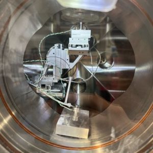
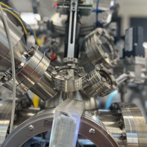
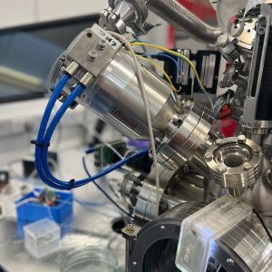
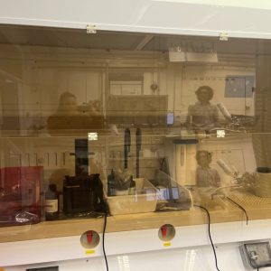
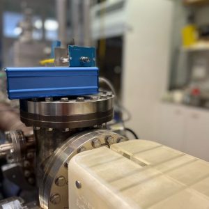
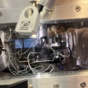
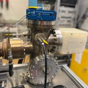
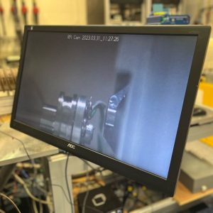
The SHeM is an atom microscope that resembles a miniaturised atom scattering beamline. Most of the system is under high (inc. sample chamber) and ultra-high vacuum and involves scattering a microscale atom beam off a sample. Samples are mounted on standard, or modified, SEM sample stubs.
The facility operates two SHeMs and the instrument used will be matched to your experimental requirements.
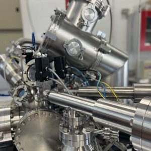
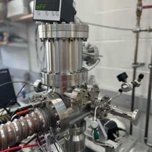
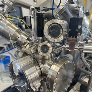
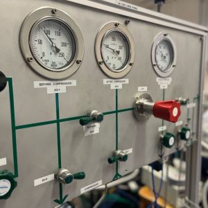


Item | Specification |
|---|---|
Beam width | 200-300nm in high resolution mode. 3-10μm in spot profile diffraction mode |
Incident beam energy | 65meV |
Sample chamber vacuum range | 1e-9 – 1e-7 mbar |
Total sample manipulation travel range | 20mm |
Depth of field | 300μm (high resolution mode), 3mm (spot profile diffraction mode) |
Angular acceptance (diffraction mode) | ~5deg |
Sample temperature range | 25C - 300C |
Samples need to be adhered to an SEM stub and must be <2cm in overall dimension. Any sample that is compatible with high vacuum may be studied. For spot profile diffraction single crystal areas of sample must be x2 larger than the beam spot size and, in most cases, should be able to withstand temperatures >100C
Experiments are unlikely to be completed in less that 1week of beam time.
Fully operational
Whether you are new to the National Facility or have visited us before, our Getting Access page contains more information about booking our services.
Please allow two weeks between booking and use.
For pricing details and more information about the services we can provide please contact us.
If you are new to CORDE we would recommend contacting us via the form below to ensure we can help you with a detailed response. Or you can contact the team directly at atom.scattering@phy.cam.ac.uk.
We’re happy to help; please contact us using the form above. Or try our searchable equipment directory for an overview of everything CORDE has to offer.